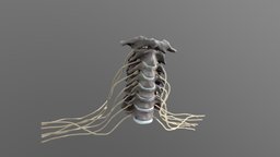
Model's Description:
Cervical spine central nervous system Anatomy 3D Model, Fully textured with UV Mapping and materials.
Download
cervical spine central nervous system Anatomy in various files formats such as Wavefront Object format, Autodesk FBX, DirectX 9.0, Stereo Lithography and HTML5 JSON format.
This is the adult cervical spine system, including C1 to C7 bones, intervertebral discs, and central nerves. I made 3D modeling based on many real anatomical photos to pursue film-level detail and fidelity - cervical spine central nervous system Anatomy - Buy Royalty Free 3D model by haibo (@haibo545001) 3D Model is ready to download for free
with 15 Texture files attached, this model contains 504200 polygons and 252051 vertices.
Model's rating is (3) based on 12 Votes.
Download : (978 Hits)
for OBJ, FBX, GLTF, GLB, DAE and other formats... or use
Model's Description:
Radial bone (radius) cat 3D Model, Optimized by RigModels.com, Fully textured with UV Mapping and materials.
Download radial bone (radius) cat in various files formats such as Wavefront Object format, Autodesk FBX, DirectX 9.0, Stereo Lithography and HTML5 JSON format.
Right radial bone (radius) of a cat
size of the specimen: 103 x 5.5 x 4 mm
3D scanning performed with the structured light scanner “Artec Micro” - radial bone (radius) cat - 3D model by vetanatMunich 3D Model is ready to download for free, this model contains 29908 polygons.
Model's Description:
Metatarsal 4 3D Model, Optimized by RigModels.com, Fully textured with UV Mapping and materials.
Download Metatarsal 4 in various files formats such as Wavefront Object format, Autodesk FBX, DirectX 9.0, Stereo Lithography and HTML5 JSON format.
3D scan of the fourth metatarsal of the left foot
Captured with Einscan Pro
Captured and edited by: Madelyn Murphy
Copyright2019 BK Alsup & GM Fox - Metatarsal 4 - Left, Unlabeled - 3D model by Bluelink Anatomy - University of Michigan (@bluelinkanatomy) 3D Model is ready to download for free, this model contains 300000 polygons.
Model's Description:
Lytic Lesions of spine 3D Model, Optimized by RigModels.com, Fully textured with UV Mapping and materials.
Download Lytic Lesions of spine in various files formats such as Wavefront Object format, Autodesk FBX, DirectX 9.0, Stereo Lithography and HTML5 JSON format.
-the vertebra is divided anatomically into the vertebral body anteriorly and the neural arch posteriorly. The neural arch is composed of bilateral pedicles, pars interarticularis, and laminae; from the neural arch arise the spinous process, bilateral transverse processes, and bilateral superior and inferior articulating processes. These anatomic components are known collectively as the posterior elements of the vertebra.
-Myeloma bone disease can result not only in fractures, but in pressure on the spinal cord (spinal cord compression), the need for surgery to prevent or repair broken bones, and/or the need for radiation treatments to the bone to control the myeloma and relieve pain. These are collectively known as “skeletal-related events.”
Reference:-
1-https://journals.lww.com/cdronline/Abstract/2017/11150/Lytic_Lesions_of_the_Posterior_Elements_of_the.1.aspx
2-https://www.myeloma.org/bone-disease - Lytic Lesions of spine - Buy Royalty Free 3D model by Ebers 3D Model is ready to download for free, this model contains 56920 polygons.
Model's Description:
Cervical Spine 3D Model, Optimized by RigModels.com, Fully textured with UV Mapping and materials.
Download Cervical Spine in various files formats such as Wavefront Object format, Autodesk FBX, DirectX 9.0, Stereo Lithography and HTML5 JSON format.
Cervical Vertebrae(C1-C7) illustrated along with spinal nerves and the vertebral arteries in the transverse foramena.
Uncinate process is labelled. Outgrowth from uncinate process(like osteophytes) can result in compression of the spinal nerve and/or vertebral arteries leading to chronic pain in the neck 3D Model is ready to download for free, this model contains 207528 polygons.


 cat")


