Model's Description:
L. Elbow with Traumatic Fusion (VCU_3D_5443) 3D Model.
Download
L. Elbow with Traumatic Fusion (VCU_3D_5443) in various files formats such as Wavefront Object format, Autodesk FBX, DirectX 9.0, Stereo Lithography and HTML5 JSON format.
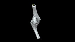
Left elbow (distal humerus and proximal ulna) with traumatic ankylosis from the American Civil War, collected by John Gouley. Specimen AFIP 1002831 from the National Museum of Health and Medicine (Silver Spring, MD). Model generated with the Segment Editor in 3D Slicer from a micro-computed tomography scan. 3D models can be downloaded from Morphosource at: https://www.morphosource.org/Detail/SpecimenDetail/Show/specimen_id/26332. Model generated by Terrie Simmons-Ehrhardt 3D Model is ready to download for free
, this model contains 1787840 polygons and 893522 vertices.
Model's rating is (3) based on 72 Votes.
Download : (968 Hits)
for OBJ, FBX, GLTF, GLB, DAE and other formats... or use
Model's Description:
Brachial bone (humerus) cat 3D Model, Optimized by RigModels.com, Fully textured with UV Mapping and materials.
Download brachial bone (humerus) cat in various files formats such as Wavefront Object format, Autodesk FBX, DirectX 9.0, Stereo Lithography and HTML5 JSON format.
Right brachial bone (humerus) of a cat
size of the specimen: 93 x 7 x 18 mm
3D scanning performed with the structured light scanner “Artec Micro” - brachial bone (humerus) cat - 3D model by vetanatMunich 3D Model is ready to download for free, this model contains 50000 polygons.
Model's Description:
Lytic humeral lesion 3D Model, Optimized by RigModels.com, Fully textured with UV Mapping and materials.
Download Lytic humeral lesion in various files formats such as Wavefront Object format, Autodesk FBX, DirectX 9.0, Stereo Lithography and HTML5 JSON format.
-When a lytic lesion is suspected, the radiologist must keep in mind the possibility of a normal variant, such as a pseudocyst. Two common locations for pseudocysts are the humeral head and the calcaneus. The pseudocyst of the humeral head is typically located in the region of the greater tuberosity, while the pseudocyst of the calcaneus is typically located anteriorly.
-Proximal humerus chondrosarcoma is a rare localization of the common primary malignant cartilaginous tumor.
Reference:
1-https://appliedradiology.com/articles/general-approach-to-lytic-bone-lesions
2-https://www.researchgate.net/figure/Proximal-humerus-lytic-lesion-with-metaphyseal-and-epiphyseal-involvement_fig1_350678996 - Lytic humeral lesion - Buy Royalty Free 3D model by Ebers 3D Model is ready to download for free, this model contains 44318 polygons.
Model's Description:
Lytic Lesions of Rib 3D Model, Optimized by RigModels.com, Fully textured with UV Mapping and materials.
Download Lytic Lesions of Rib in various files formats such as Wavefront Object format, Autodesk FBX, DirectX 9.0, Stereo Lithography and HTML5 JSON format.
-In skeletal tumors, metastatic bone tumors have the most common occurrence [1] and primary bone tumors of chest wall accounts for only 5 - 8 % of all bony tumors.
-Of these approximately 95% occur in ribs and remainder in the sternum.
Reference :-
https://www.fortunejournals.com/articles/expansile-lytic-lesions-of-rib-two-rare-case-reports.html - Lytic Lesions of Rib - Buy Royalty Free 3D model by Ebers 3D Model is ready to download for free, this model contains 118150 polygons.
Model's Description:
Left Frontosphenoidal Craniosynostosis 3D Model.
Download Left Frontosphenoidal Craniosynostosis in various files formats such as Wavefront Object format, Autodesk FBX, DirectX 9.0, Stereo Lithography and HTML5 JSON format.
An infant cranium and mandible (6 months old) with left frontosphenoidal craniosynostosis. Small gaps between cranial bones were filled to make it easier to 3D print. The mandible was also solidified to make 3D printing easier (unerupted teeth are not visible)–central mandibular incisors are erupted. You can also cut the cranium before 3D printing to have access to the endocranial anatomy.
I segmented this from a computed tomography scan from Embodi3D.
Download includes beige PLY of the cranium and mandible combined. The additional file contains the separated STL models for 3D printing 3D Model is ready to download for free, this model contains 339948 polygons.
 3D Model")

 cat")


